


Mortons Neuroma
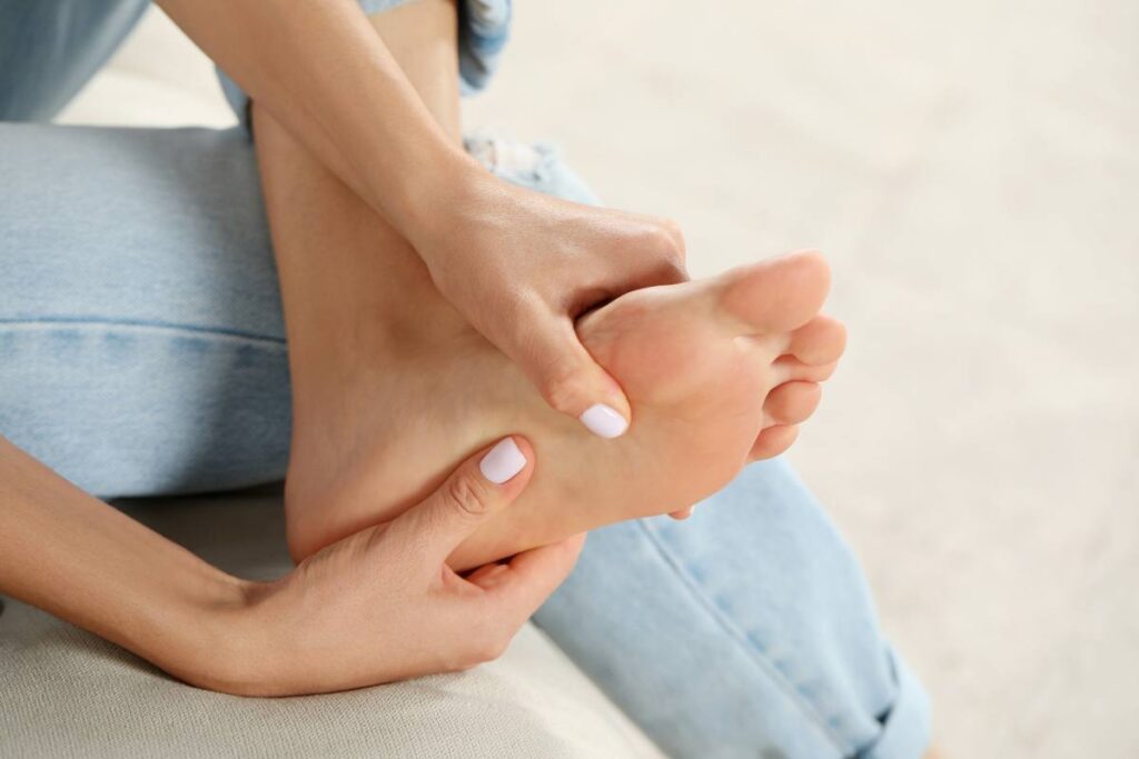
Morton’s neuroma is a common cause of pain in the ball of the foot, usually felt between the toes. It happens when one of the small nerves in this area becomes irritated and swollen, often due to pressure from footwear or activities that place extra load on the front of the foot.
People often describe the sensation as sharp or burning pain, or as though they are standing on a small pebble. Symptoms may come and go and can feel worse when wearing tight shoes or doing activities that involve a lot of walking or running. The good news is that with the right advice and changes to footwear or activity, symptoms often improve. (1,2)
Frequently Asked Questions
Morton’s neuroma is a condition where a nerve in the front of the foot becomes irritated, usually between the third and fourth toes. This can lead to sharp, burning pain or a feeling like there’s a small pebble under your foot when walking or wearing tight shoes.
The estimated prevalence is around 1% in the general population.
Although it’s a relatively common cause of forefoot pain, with it being present in up to 1 in 3 patients with forefoot symptoms. (7)
No. Although painful, Morton’s neuroma is not a sign of anything else significantly occurring or any links to other health conditions.
Symptoms can worsen if aggravating load isn’t modified with early conservative management highly effective. (1)
More common in women, with a female-to-male ratio of 4:1.
Typical symptoms to occur people aged between 40–60.
Risk factors include tight wearing footwear and repetitive impact on the forefoot. (2)
Sharp, burning forefoot pain.
Numbness or tingling into adjacent toes.
Sensation of “walking on a pebble” or a “marble”. (7)
Modify footwear (wide toe box; avoid heels).
Use cushioned/forefoot supporting insoles.
Reduce forefoot load; consider activity modification.
Trial NSAIDs/ice for symptom relief. (1)
Recovery timescale can be variable.
Many patients improve over weeks to months with conservative management.
Persistent cases may require injection or (rarely) surgery. (1)
We recommend consulting a musculoskeletal physiotherapist to ensure exercises are best suited to your recovery. If you are carrying out an exercise regime without consulting a healthcare professional, you do so at your own risk.

Morton’s neuroma typically presents with burning, sharp pain in the ball of the foot, often between the 3rd and 4th toes.
Frequently, paraesthesia, numbness or a distinctive sensation of stepping on a pebble is also a symptom.
Symptoms may be intermittent, triggered by forefoot loading, narrow footwear or prolonged standing and are sometimes relieved by removing shoes. (2,4)
The condition results from repetitive compression or irritation of the common digital plantar nerve, most commonly in the intermetatarsal spaces (between the long bones of the foot).
Long term mechanical stress, hyperextension of the toe joints, tight footwear and anatomical factors such as foot posture variations can contribute to inflammation and nerve irritation. (3,7)
This is not an exhaustive list. These factors could increase the likelihood of someone developing Morton’s neuroma. It does not mean everyone with these risk factors will develop symptoms.
Common risk factors include:

Morton’s neuroma affects approximately 1% of the general population but occurs more frequently in people with general forefoot pain. Women are 4 times more likely to develop the condition, with peak incidence between ages 40 and 60. Bilateral (both feet) involvement can occur in up to 15–21% of cases. (2,7)
Musculoskeletal physiotherapists and other appropriately qualified healthcare professionals can provide you with a diagnosis by obtaining a detailed history of your symptoms. A series of physical tests might be performed as part of your assessment to rule out other potentially involved structures and gain a greater understanding of your physical abilities to help facilitate an accurate working diagnosis.
Your treating clinician will want to know how your condition affects you day-to-day so that treatment can be tailored to your needs and personalised goals can be established. Intermittent reassessment will ascertain if you are making progress towards your goals and will allow appropriate adjustments to your treatment to be made. Imaging studies like ultrasound scans can be arranged if conservative management does not settle symptoms. (2)
Diagnosis is primarily clinical. Key features include:
Imaging is useful when diagnosis is unclear or for organising further care:
Although imaging can be organised, clinical assessment alone has demonstrated approximately 96% sensitivity for correct diagnosis. (4,8)
As part of the sessions with your physiotherapist, they will help you to understand your condition and what you need to do to help the recovery from your Morton’s neuroma. This may include reducing the amount or type of activity, as well as other advice aimed at reducing your pain. It is important that you try and complete the exercises you are provided as regularly as possible to help with your recovery. Rehabilitation exercises are not always a quick fix, but if done consistently over weeks and months then they will, in most cases, make a significant difference.
Strategies that may help include:
These approaches often reduce symptoms and help avoid progression. (1)
In some instances, a one-to-one assessment is appropriate to individually tailor targeted rehabilitation.
Our team of expert musculoskeletal physiotherapist can create rehabilitation plans to enable people to manage their condition. If you have any questions or concerns about a condition, we recommend you book an consultation with one of our clinicians.
Most patients can return to normal activity once symptoms are controlled and forefoot load is managed effectively. Gradual loading, supportive footwear and symptom monitoring are essential. Chronic cases may require periods of de–loading or orthotic support before resuming higher impact sports. (1,7)
If conservative management fails, additional treatments include:
References
Other Conditions in Feet